
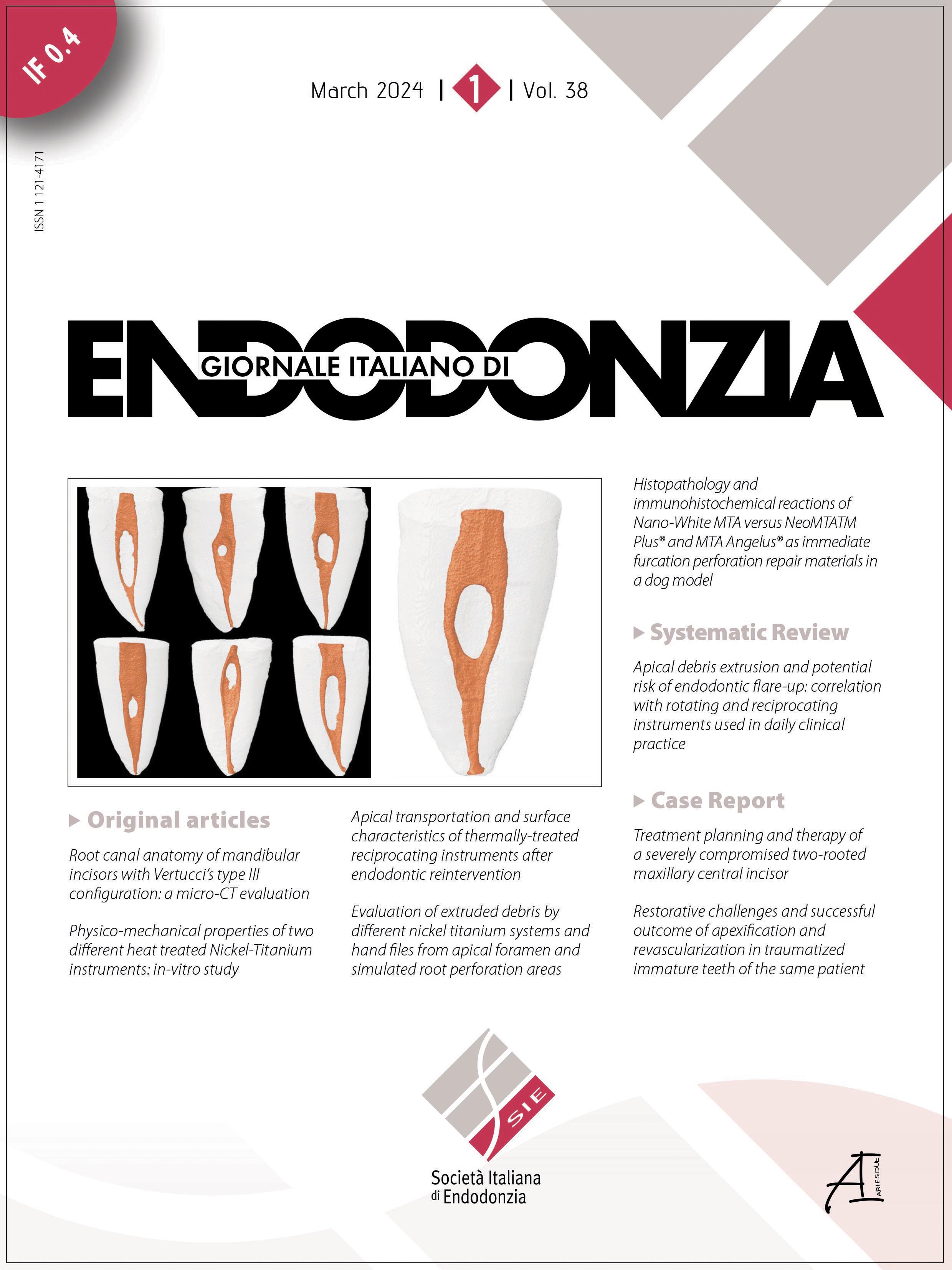
Root canal treatment of a maxillary first molar with unusual anatomy: a case report

All claims expressed in this article are solely those of the authors and do not necessarily represent those of their affiliated organizations, or those of the publisher, the editors and the reviewers. Any product that may be evaluated in this article or claim that may be made by its manufacturer is not guaranteed or endorsed by the publisher.
Accepted: 13 December 2023
Authors
Aim: To describe the management of a maxillary first molar with six root canals, two in each root.
Summary: A 36-year-old male patient presented with a complaint of discoloration and decay on an upper left back tooth. Clinical examination revealed the presence of deep occlusal caries on the maxillary left first molar. A detailed clinical and radiographical examination led to a diagnosis of pulp necrosis with asymptomatic apical periodontitis. Non-surgical root canal treatment was performed. Due to the suspected anatomical variation, Cone beam computed tomography (CBCT) imaging was obtained. CBCT imaging and clinical identification using the DOM revealed the presence of six root canals, two in each root, with the code (326 MB2 DB2 P2-1) using Ahmed et al. system. Root canal treatment was successfully performed and confirmed radiographically. The tooth was restored with a resin composite restoration. The patient was referred for prosthodontic evaluation for extracoronal restoration.
Key learning points:
- This case highlights how clinicians must approach root canal treatment with a comprehensive mindset that considers the potential for extraordinary anatomical complexities.
- Preoperative knowledge of root canal anatomy and proper armamentarium are mandatory for successful root canal treatment procedures.
Downloads
Citations


How to Cite

This work is licensed under a Creative Commons Attribution-NonCommercial 4.0 International License.