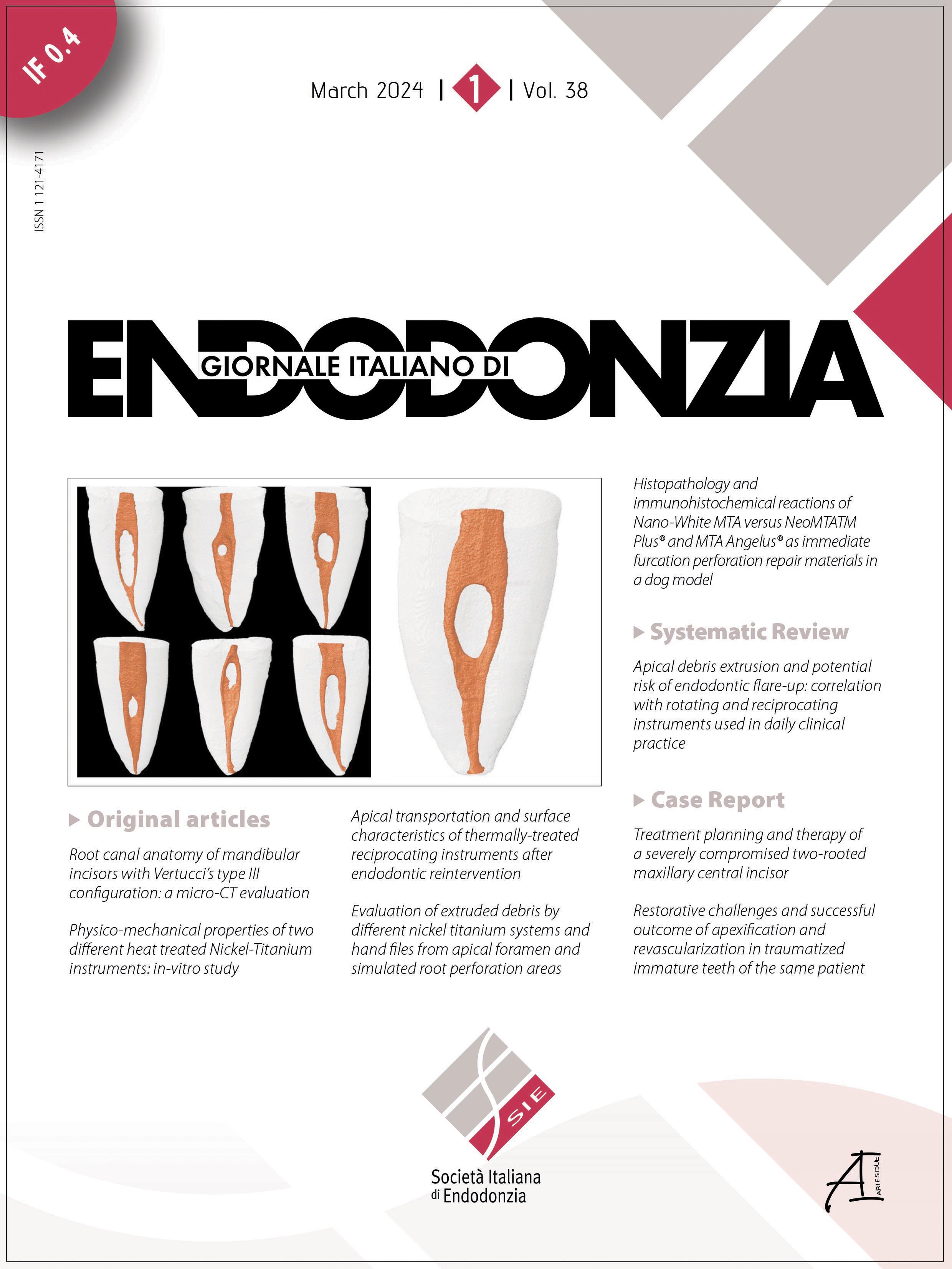
Histopathology and immunohistochemical reactions of Nano-White MTA vs. NeoMTATM Plus® and MTA Angelus® as immediate furcation perforation repair materials in a dog model


Submitted: 9 September 2023
Accepted: 29 October 2023
Published: 16 November 2023
Abstract Views: 407
PDF: 64
Publisher's note
All claims expressed in this article are solely those of the authors and do not necessarily represent those of their affiliated organizations, or those of the publisher, the editors and the reviewers. Any product that may be evaluated in this article or claim that may be made by its manufacturer is not guaranteed or endorsed by the publisher.

Authors

Aim: This study compared histopathology and immunohistochemical reactions of Nano-white mineral trioxide aggregate (Nano-WMTA), Neo-mineral trioxide aggregate plus (NeoMTATM Plus®), and mineral trioxide aggregate angelus (MTA Angelus®) as furcation perforation repair materials.

Methodology: Twelve premolars were used in six mongrel dogs. Seventy two teeth were divided according to post-treatment evaluation period into two main equal groups (three dogs' each/36 teeth each) including; group I: after one month and group II: after three months. Each main group was subdivided into three experimental subgroups (8 teeth each) according to the immediate perforation repair materials used, and two control groups. The perforations in subgroups 1, 2 and 3 were sealed with NeoMTATM Plus®, Nano-WMTA and MTA Angelus®, respectively. In the positive control subgroup (8 teeth), the perforations were done with no repair. Negative control subgroup (4 teeth) represented intact teeth with no perforation to show the normal histology. Pulpotomy and root canal obturation were carried out in the experimental and positive control subgroups. Inflammatory cell count and new hard tissue formation were evaluated using histopathology and immunohistochemical staining with Osteonectin. All data were statistically analyzed.

Results: All tested materials demonstrated significant lower inflammatory cell counts than the positive control subgroup and significant higher new bone formation than both positive and negative subgroups. There was no statistically significant difference in inflammatory cell count and new hard tissue formation among the experimental materials in both groups (p<0.05).

 Conclusion: All tested materials effectively reduced inflammation and promoted hard tissue formation with no statistically significant difference.


Supporting Agencies

None

Elkhashab, R., Mahran, A., Badr, M., & Abu-Seida, A. (2023). Histopathology and immunohistochemical reactions of Nano-White MTA vs. NeoMTA<sup>TM</sup> Plus<sup>®</sup> and MTA Angelus<sup>®</sup> as immediate furcation perforation repair materials in a dog model. Giornale Italiano Di Endodonzia, 38(1). https://doi.org/10.32067/GIE.2023.37.01.27

Downloads

Download data is not yet available.

Citations