
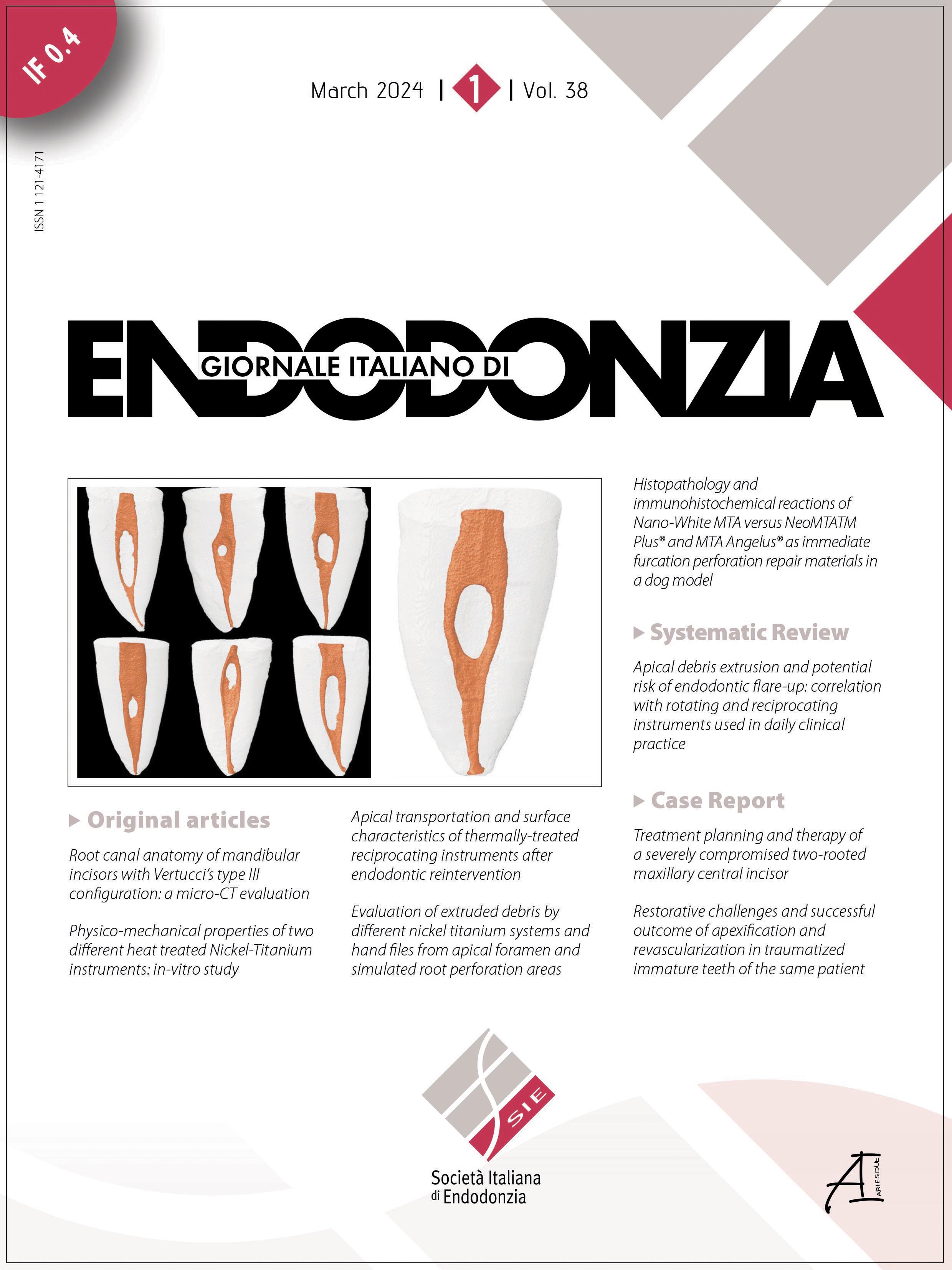
Root canal anatomy of mandibular incisors with Vertucci’s type III configuration: a micro-CT study

All claims expressed in this article are solely those of the authors and do not necessarily represent those of their affiliated organizations, or those of the publisher, the editors and the reviewers. Any product that may be evaluated in this article or claim that may be made by its manufacturer is not guaranteed or endorsed by the publisher.
Accepted: 30 November 2023
Authors
Aim: To evaluate the root canal anatomy of mandibular incisors with Vertucci’s type III configuration.
Methodology: Forty mandibular incisors were scanned using a microCT to measure canal and dentin volume, bifurcation and merging levels, minor and major diameters, long-short diameter ratio, dentin thickness, degree of curvatures and number of foramens.
Results: The apical third showed lower volume of canal and dentin. The bifurcation with the formation of buccal and lingual canals presented a mean of 3.75 mm extension. The cement-enamel junction, bifurcation and merging levels showed major diameter (P < 0.05). The round shaped canals were found in buccal (67.5%), lingual (85%) and apical sections (55%). In apical section dentin thickness ranged from 1.02 mm to 0.52 mm. No specimen showed root curvature and 82.5% of mandibular incisors presented single apical foramen.
Conclusion: The morphologic aspects of root canal bifurcation and merging in mandibular incisors Vertucci’s type III do not present a consistent pattern.
Downloads
Citations


Supporting Agencies
Brazilian National Council for Scientific and Technological DevelopmentHow to Cite

This work is licensed under a Creative Commons Attribution-NonCommercial 4.0 International License.